Our 3D-EXplore Tumoroid Technology
Build a detailed, highly nuanced understanding of the effect your candidate therapeutic exerts on patient tumor tissue, including the tumor microenvironment (TME), with Nilogen’s proprietary 3D-EXplore tumoroid technology.
A TECHNOLOGY UNLIKE ANY OTHER
While there are many different types of 3D cell technologies, Nilogen’s unique 3D-EXplore tumoroid platform provides the most clinically relevant model of patient tumor biology available.
By minimally processing fresh patient tumor tissue into uniformly sized tumoroids and pooling tumoroids before aliquoting into individual wells, we create highly reproducible representations of the complete tumor in multiple assay wells.
The result is a platform that enables replicate studies and/or integratable analysis by multiple methods—think high content imaging and gene expression analysis and multiplex cytokine analysis and multi-parameter flow cytometry —and delivers similar treatment response rates to that seen in patients.

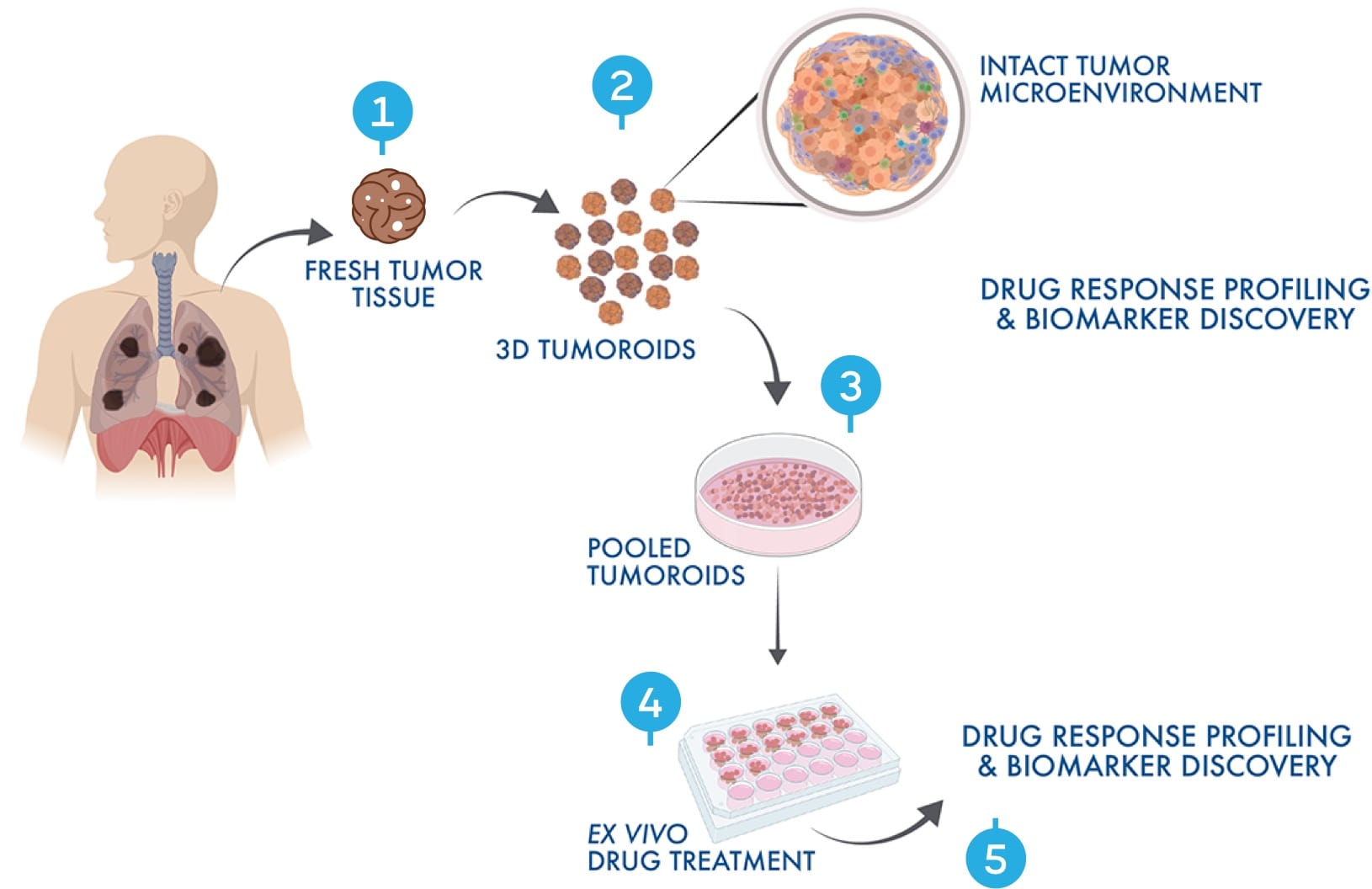
HOW OUR 3D TUMOROID TECHNOLOGY WORKS
Each well delivers highly reproducible representations of the entire heterogeneous tumor complete with intact tumor microenvironment (TME).
-
ISOLATION OF FRESH TUMOR TISSUE
We start with freshly isolated tissue ethically sourced from our extensive network of clinics in the US.
-
CONVERSION INTO 3D TUMOROIDS
Each fresh tumor is minimally processed using a proprietary mechanical method into uniformly-sized tumoroids of about 150 µm in diameter. No chemical dissociation, propagation or reassembly.
-
POOLING AND ALIQUOTING OF TUMOROIDS
To account for the heterogeneity that exists within each tumor, we pool up to 9,000 tumoroids from a single tumor before aliquoting hundreds of tumoroids into individual wells of a multi-well plate, minimising well-to-well variability.
-
EX VIVO TREATMENT WITH CANDIDATE THERAPIES
A single tumor can provide enough tumoroids for many study arms with multiple assays per arm, which are then exposed to drug/combinations. Each drug/combination can be assessed using multiple analytical techniques, resulting in a comprehensive, multi-omics view of multiple drugs/combinations in each tumor.
-
DRUG RESPONSE PROFILING & BIOMARKER DISCOVERY
Nilogen uses sophisticated bioinformatics and artificial intelligence algorithms to analyze and integrate your data, ultimately delivering a fully analyzed, interpreted, and actionable report of your study.
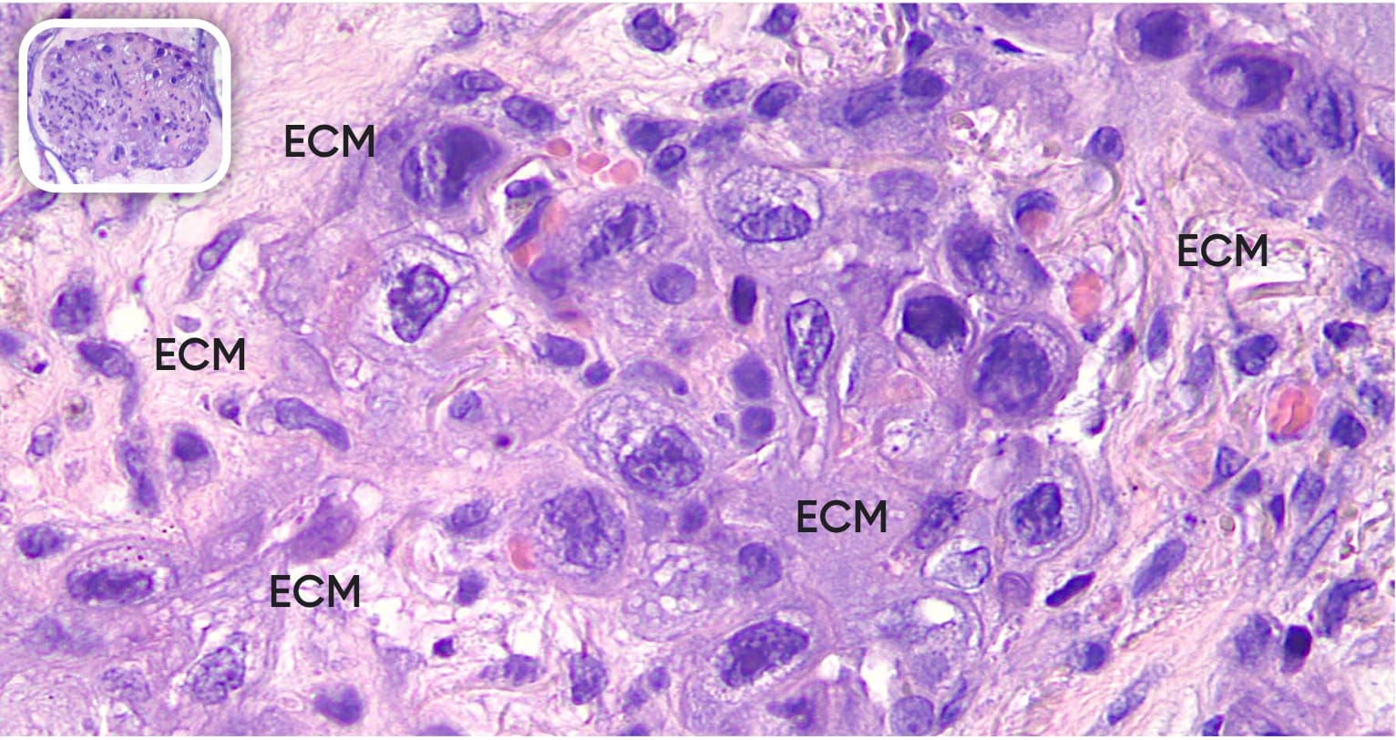
SEE THE DIFFERENCE IN NILOGEN’S TUMOROIDS
In this example of a pancreatic tumoroid, the extracellular matrix (ECM) is preserved in addition to cell-cell and cell-ECM interactions.






Frequently asked questions
Nilogen derives its tumoroids from fresh, never frozen patient tumor tissue; the tumoroids are generated within 24 hours of the resection of the tumor. The mechanism used to generate the tumoroids is a purely mechanical process that does not use any sort of chemical digestion and produces tumoroids of uniform size and shape. There are no cell propagation steps, nor any reassembly steps. This method allows for the capture of the unique heterogeneity within each individual tumor from a structural, morphological, and phenotypic standpoint. Additionally, the stromal environment and the communication that occurs from cell-cell and cell-extracellular matrix interactions remains intact.
No, only tumoroids derived from a single tumor are pooled. This allows the heterogeneity of the individual tumor to be captured while being able to create a homogenous population of tumoroids to be distributed from well to well for different assays and treatment arms.
The number of assays and treatment arms that can be performed on one tumor depends on a number of variables including: the tumor type, the assays to be run, and the number of timepoints to be sampled. Most tumors (i.e., non small lung cancer, renal cell carcinoma, colorectal cancer) yield 9,000 tumoroids. Different assays also require different numbers of tumoroids. As an example, 400 tumoroids are needed for each treatment arm at each timepoint for 1 flow cytometry panel (the same number are required for RNA sequencing), whereas only 100 tumoroids are needed for our tumor cell killing assay. The complexity of your study dictates how many arms can be run on each tissue, but even a complex set of assays can support 10 arms of more.
To ensure that tumoroids have functional tumor resident lymphoid cells that respond to external stimuli we use phorbol 12-myristate 13-acetate plus ionomycin (PMAI) and or CD3/CD28 mAbs as positive controls in our ex vivo assays.
Featured Applications
GET STARTED WITH NILOGEN
Find out how Nilogen’s 3D-EXplore tumoroid technology can advance your oncology program.
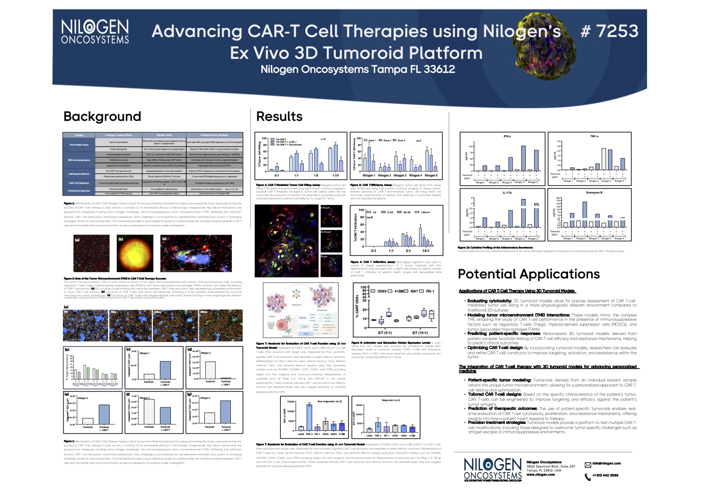
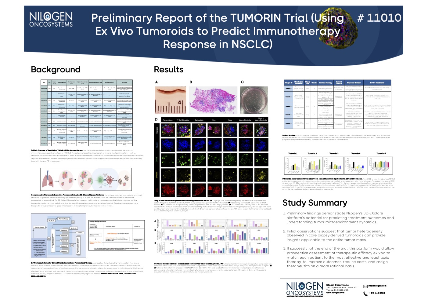
Nilogen Posters
See the robustness of our 3D tumoroid technology in our recent conference posters.
