Tumor Killing Assay
The tumor microenvironment consists of multiple cell types including tumor cells, immune cells and stroma. Nilogen’s high-content 3D imaging system is capable of identifying tumor and non-tumor cells during our tumor killing assay and our proprietary analytics enables the quantitative analysis of cell killing in response to drug treatment.
WHY IS TUMOR CELL KILLING IMPORTANT?
Cytotoxic T cells (CTL) are key components of the adaptive immune response and natural killer (NK) cells play an analogous role in the innate immune response in combating tumors. Tumors exhibit a variety of mechanisms to evade the immune system; using our advanced tumor killing assay, allows us to develop novel approaches to augment anti-tumor immunotherapy and cytolytic killing by both NK and cytotoxic T cells. Nilogen’s 3D-EXplore platform utilizes
Capabilities
High Content Confocal Microscopy
Quantify and match the ability of your drug to kill tumor cells with the expression of your target antigen, induce phagocytosis and measure penetration into the tumor microenvironment. Evaluate in conjunction with other proteogenomic assays to link mechanisms of action with effect.
Flow Cytometry
Nilogen’s extensive experience in multiparameter flow cytometry matched with our optimized panels enable our clients to evaluate the immune landscape, T-cell activation, checkpoint inhibition, ADCCs, ADC, myeloid function, phagocytosis and cellular proliferation as well as develop and deploy custom panels.
TOTALSeq
TOTALSeq works hand-in-hand with scRNAseq to enable the simultaneous measurement of protein and RNA at the single cell level, providing enhanced cell type identification. Evaluate the impact of drug therapy on up to 130 protein markers in conjunction with the transcriptome of each cell; this enables the single cell analysis of individual immune cells in a heterogeneous tumor microenvironment.
Multiplex Immunohistochemistry
Multiplex Immunohistochemistry/Immunofluorescence (mIHC/IF) provides high-throughput multiplex staining and standardized semi-quantitative analysis for highly reproducible, efficient and cost-effective tissue studies. This technique allows the simultaneous detection of multiple markers on a single tissue section, providing a comprehensive view of tissue composition, pre- and post-treatment.
Multiplex Cytokine Analysis
We enable the simultaneous analysis of up to 100 cytokines and chemokines. Traditional disassociated 2D models and organoids lack an intact tumor microenvironment and disrupt the signals crucial to tumorigenesis and drug response. Nilogen’s 3D-EXplore tumoroids preserve these intricate interactions. At Nilogen we provide a broad range of cytokine assay services including single plex up to 100-plex panels for multiplex cytokine analysis to assess immune activity and drug mediated responses.Our cytokine profiling service provides an enhanced approach to study mechanisms of action in living tumor tissue.
Scientific Data
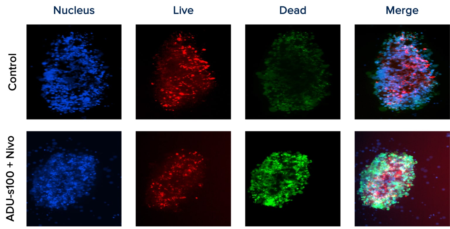
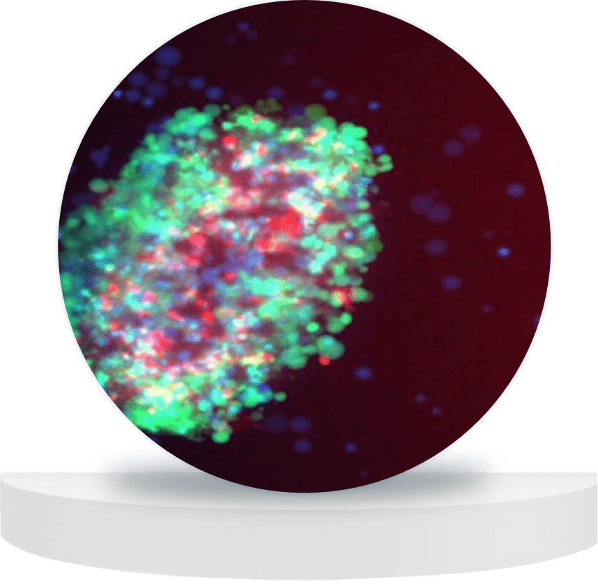
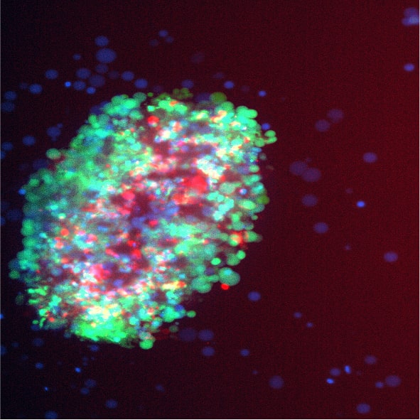
Nilogen's tumor cell killing assay was performed using high content confocal imaging to visualize treatment-mediated changes in the viability of tumor cells within live tumoroids. Images show increased tumor cell death with the combination treatment of ADU-S100 + Nivolumab as compared to controls.
Related Resources
Browse our latest posters and presentations using Nilogen's fresh patient tumoroid technology.
