Cell-Based Immunotherapies
3D-EXplore can be used to investigate and qualify the penetration, and tumor cell killing capabilities of all cell-based immunotherapies including Tumor Infiltrating Lymphocyte (TIL) therapy, engineered T cell receptor (TCR) therapy, Gamma delta (γδ T-cells ) cell therapy and chimeric antigen receptor T cell (CAR-T) therapy.
WHY ARE CELL-BASED IMMUNOTHERAPIES IMPORTANT?
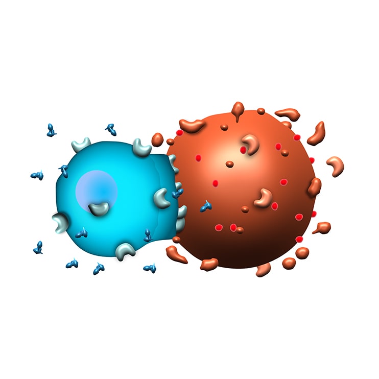
Adoptive cell therapies (ACTs), also known as cellular or cell-based immunotherapies, are therapeutic interventions that utilize immune cells to target tumor cells. Cytotoxic T cells are particularly powerful against cancer, due to their ability to bind to antigens on the surface of cancer cells. Cell-based immunotherapies take advantage of this natural ability, and with 3D-EXplore we have the unique ability to investigate and quantify the penetration and efficacy of these therapies.
Capabilities
Flow Cytometry
Nilogen’s extensive experience in flow cytometry matched with our optimized panels enable our clients to evaluate the immune landscape, T-cell activation, checkpoint inhibition, ADCC, myeloid function, phagocytosis and cellular proliferation as well as develop and deploy custom panels.
High Content Confocal Microscopy
Quantify and match the ability of your drug to kill tumor cells with the expression of your target antigen, induce phagocytosis and measure penetration into the tumor microenvironment. Evaluate in conjunction with other proteogenomic assays to link mechanisms of action with effect.
Single Cell RNA Sequencing
Enables the quantitative measurement of molecular activity that underlies the phenotypic diversity of cells within a tumor by sequencing individual cells. Intratumor heterogeneity is common across all tumor types; accurate characterization of this heterogeneity is essential for determining the mechanisms of cancer pathogenesis and identifying novel targets for immunotherapy and drug development.
TOTALSeq
TOTALSeq works hand-in-hand with scRNAseq to enable the simultaneous measurement of protein and RNA at the single cell level, providing enhanced cell phenotyping. Evaluating the impact of drug therapy on up to 130 protein markers in conjunction with the transcriptome of each cell, enables single cell analysis of individual immune cells in a heterogeneous tumor microenvironment.
Multiplex Immunofluorescence
Multiplex Immunofluorescence (IF) provides high-throughput multiplex staining and standardized quantitative analysis for highly reproducible, efficient and cost-effective tissue studies. This technique allows the simultaneous detection of multiple markers on a single tissue section, providing a comprehensive view of tissue composition and spatial relationships, pre- and post-treatment.
SCIENTIFIC DATA
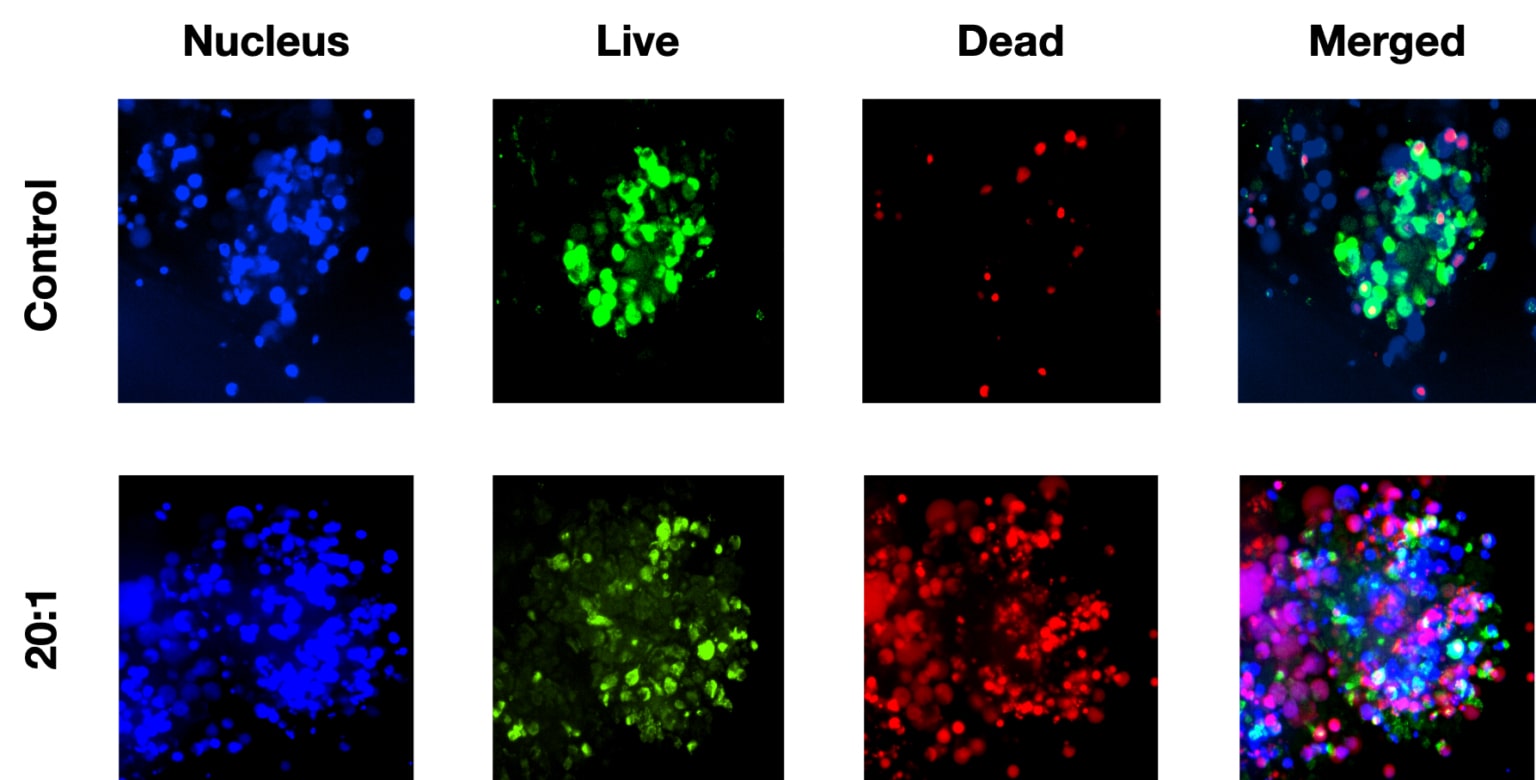
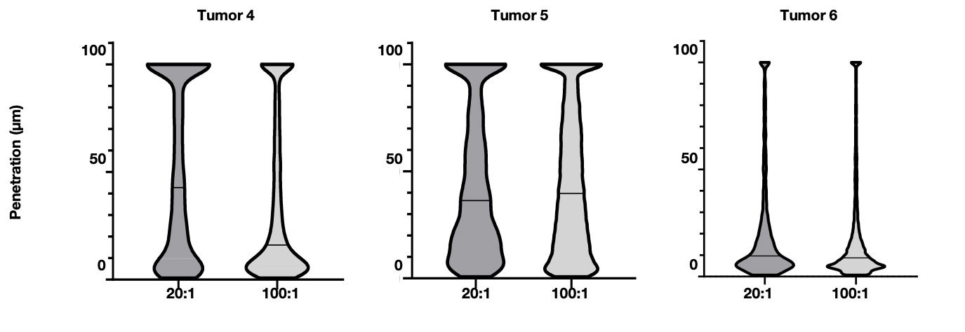
High content confocal microscopy can be used to quantify tumor-mediated cell killing as shown in this example image. Comparing the number of dead cells following treatment with CAR-T cells in control versus treated tumoroids, as well as quantify the penetration distance of cell tracker CAR-Ts. This demonstrates that the heterogeneity of the tumor microenvironment heavily influences the effectiveness of the therapy with varying levels of penetration and CAR-T-mediated tumor cell killing due to the unique stromal composition of each tumor.
Related Resources
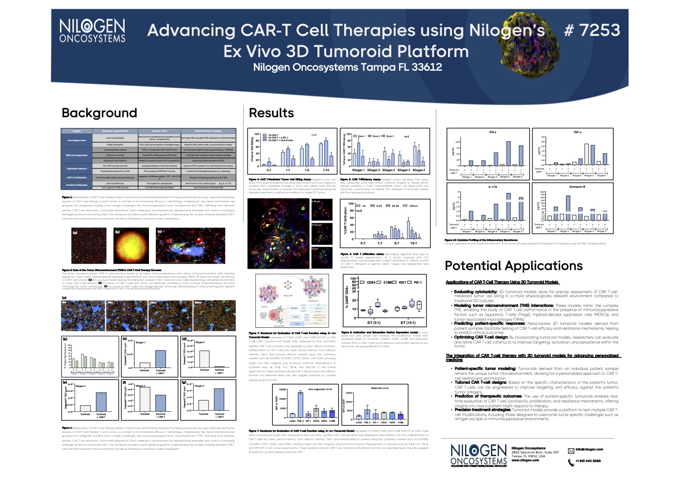
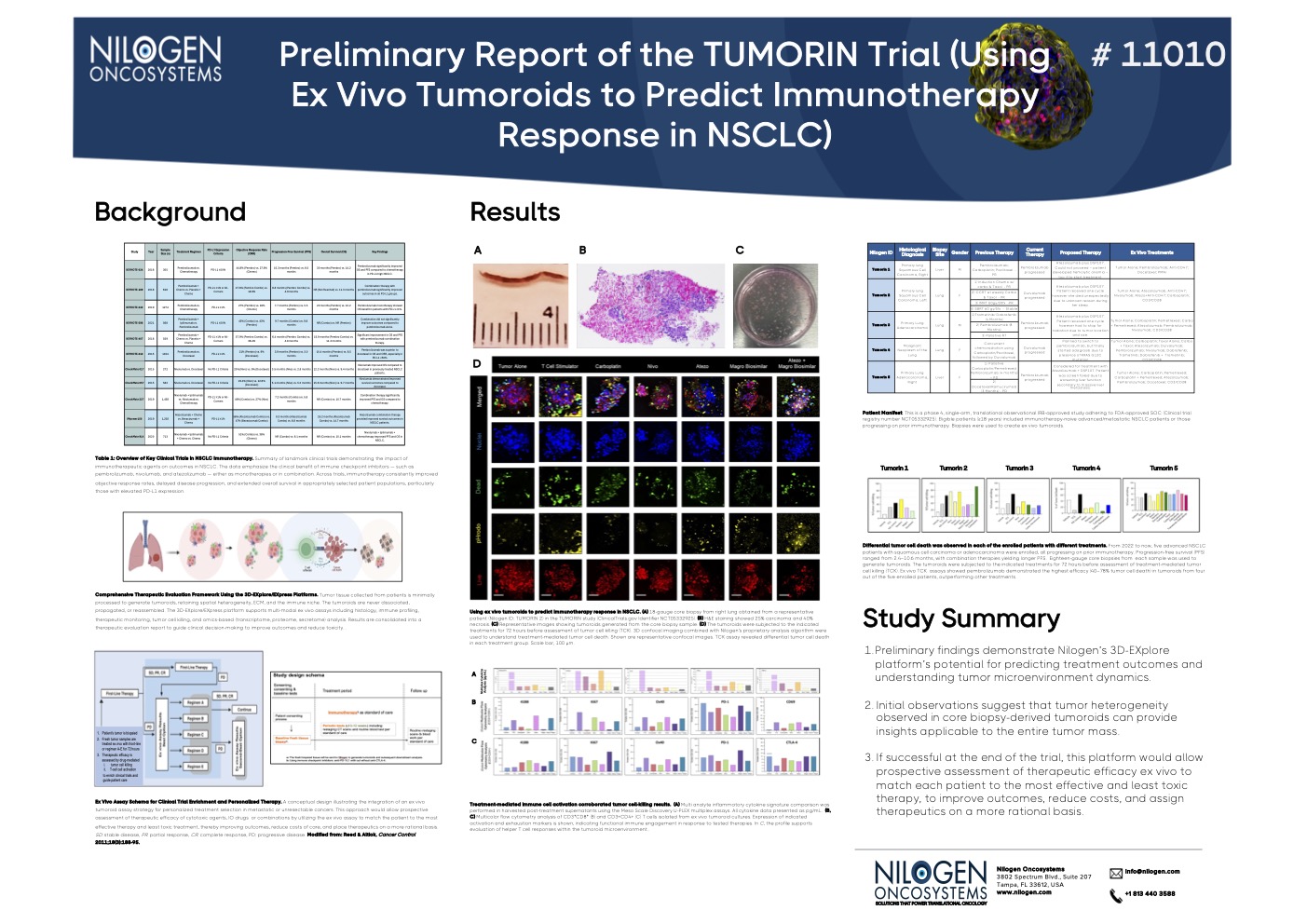
Browse our latest posters and presentations using Nilogen's fresh patient tumoroid technology.

